Adenine, thymine, cytosine, and guanine represent the four nitrogen bases shown in model 1, forming the chemical alphabet that stores and transmits genetic information across all living organisms. These molecular units function like letters in a biological language, combining in precise sequences to encode instructions for growth, repair, and reproduction. When students examine model 1, they observe how these bases pair and stack to create the elegant architecture of nucleic acids, revealing why heredity remains both stable and adaptable. Understanding these four nitrogen bases allows learners to visualize how microscopic components build macroscopic life, connecting chemistry to biology in a seamless narrative that supports deeper inquiry into genetics, medicine, and biotechnology Surprisingly effective..
It sounds simple, but the gap is usually here Simple, but easy to overlook..
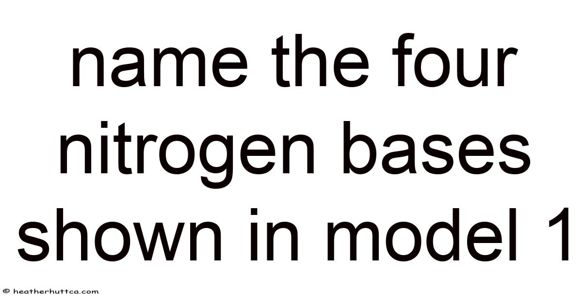
Introduction to Nitrogen Bases in Model 1
Model 1 introduces learners to the molecular foundation of genetic material by displaying the four nitrogen bases that compose DNA. Still, these compounds earn their name because each contains nitrogen atoms within ring-shaped structures that participate in hydrogen bonding and molecular recognition. Which means in the context of model 1, the bases appear as distinct shapes and colors, helping students differentiate chemical identities while recognizing patterns of complementarity. By focusing on these units, educators stress that genetic information does not emerge from abstract forces but from tangible chemical interactions. The clarity provided by model 1 supports accurate mental models, enabling students to predict how changes at the base level influence higher-order biological outcomes Simple as that..
The Four Nitrogen Bases Shown in Model 1
When examining model 1, four nitrogen bases stand out through their structural differences and functional roles. Each belongs to one of two chemical classes, which determines how it pairs with a partner across the double helix Turns out it matters..
Purines: Adenine and Guanine
Purines consist of two fused rings containing both carbon and nitrogen atoms, giving them a larger profile compared to their counterparts. In model 1, adenine and guanine appear as broader structures that fit precisely into complementary spaces along the nucleic acid chain.
- Adenine pairs with thymine through two hydrogen bonds, creating a stable but reversible connection that supports accurate replication and transcription.
- Guanine pairs with cytosine through three hydrogen bonds, producing a slightly stronger interaction that contributes to the overall stability of the double helix.
These pairing rules, clearly illustrated in model 1, make sure genetic information remains faithful during cell division while allowing controlled separation when proteins need to access the code.
Pyrimidines: Thymine and Cytosine
Pyrimidines consist of a single ring structure, making them smaller than purines. In model 1, thymine and cytosine display compact shapes that reflect their chemical simplicity and specific bonding capabilities.
- Thymine accepts two hydrogen bonds from adenine, maintaining consistent spacing between the two DNA strands.
- Cytosine accepts three hydrogen bonds from guanine, reinforcing structural integrity and supporting error-checking mechanisms during DNA synthesis.
The regularity of these base pairings, as demonstrated in model 1, explains why DNA maintains a uniform width despite variations in sequence.
Structural Features Highlighted in Model 1
Model 1 does more than label the four nitrogen bases; it reveals how their shapes and chemical properties produce a functional whole. By representing hydrogen bond donors and acceptors, the model clarifies why adenine cannot stably pair with cytosine or guanine, and why thymine and cytosine maintain their distinct identities. This specificity arises from the arrangement of nitrogen and oxygen atoms that create regions of partial positive and negative charge, encouraging selective attraction That alone is useful..
Additionally, model 1 illustrates how the bases align in a stacked arrangement, with flat ring systems positioned one above another. Even so, this stacking generates hydrophobic interactions that exclude water from the interior of the double helix, stabilizing the molecule in the aqueous environment of the cell. Students who study model 1 gain insight into how chemistry and physics cooperate to produce a genetic system capable of both durability and controlled flexibility Worth keeping that in mind..
Functional Significance of the Four Nitrogen Bases
The four nitrogen bases shown in model 1 serve as more than structural components; they act as carriers of biological meaning. The sequence of these bases along a DNA strand constitutes a code that determines which proteins a cell will produce and when. During replication, enzymes read the base sequence and synthesize a complementary strand, ensuring that each daughter cell inherits an accurate copy of the genetic blueprint Small thing, real impact..
In transcription, the base sequence guides the synthesis of RNA molecules that transport instructions to ribosomes, where proteins are assembled. On the flip side, errors in base pairing can lead to mutations, some of which may alter protein function or regulation. By understanding how model 1 represents correct and incorrect pairings, learners appreciate the precision required for life to persist across generations Easy to understand, harder to ignore..
Scientific Explanation of Base Pairing and Stability
The stability of DNA arises from a combination of hydrogen bonding and base stacking, both of which are evident in model 1. Hydrogen bonds form between electronegative atoms and hydrogen atoms bonded to nitrogen, creating reversible links that allow the strands to separate without breaking covalent bonds. Although individually weak, these bonds collectively contribute significant stability when multiplied across millions of base pairs.
It sounds simple, but the gap is usually here.
Base stacking, meanwhile, results from interactions between the electron clouds of adjacent ring systems. Now, these forces, known as London dispersion forces, strengthen the double helix by minimizing exposure of hydrophobic surfaces to water. Model 1 helps students visualize how the regular spacing and planar shapes of the four nitrogen bases optimize these interactions, producing a molecule that is both flexible and resilient Worth keeping that in mind. Worth knowing..
Educational Value of Model 1
Model 1 serves as a bridge between abstract concepts and tangible understanding. By displaying the four nitrogen bases in a clear, organized format, it reduces cognitive load and supports pattern recognition. On top of that, students can trace the path of information from gene to protein, seeing how chemical properties translate into biological outcomes. This visual approach benefits diverse learners, including those who struggle with text-heavy explanations but excel when presented with structured diagrams Worth knowing..
Worth adding, model 1 encourages inquiry-based learning by prompting questions about mutation, repair, and evolution. When students ask why certain pairings occur or how errors are corrected, they engage in scientific reasoning that deepens their mastery of genetics. The model thus functions not only as a teaching tool but also as a catalyst for curiosity and critical thinking Worth keeping that in mind. No workaround needed..
Counterintuitive, but true.
Common Misconceptions Clarified by Model 1
Despite its clarity, model 1 can be misinterpreted if students overlook key details. While guanine-cytosine pairs involve three hydrogen bonds and adenine-thymine pairs involve two, covalent bonds within the sugar-phosphate backbone remain far stronger than any inter-base interaction. One common misconception involves confusing the number of hydrogen bonds with bond strength in a chemical sense. Model 1 helps distinguish these bond types, reinforcing that DNA strands separate not because hydrogen bonds break under force, but because enzymes actively unzip the helix in a controlled manner.
Another misconception concerns the universality of thymine. That's why in DNA, thymine is indeed one of the four nitrogen bases shown in model 1, but in RNA, thymine is replaced by uracil. By comparing model 1 with representations of RNA, learners can appreciate how small chemical changes produce significant functional differences, such as increased stability in DNA for long-term storage versus flexibility in RNA for transient messaging The details matter here..
Conclusion
The four nitrogen bases shown in model 1 — adenine, thymine, cytosine, and guanine — form a molecular language that encodes the instructions for life. Through precise pairing, strategic stacking, and selective bonding, these bases create a genetic system capable of stability, replication, and controlled variation. Model 1 illuminates these principles with clarity, enabling students to visualize how chemistry underlies biology and how structure determines function. By mastering the details presented in model 1, learners build a foundation for exploring genetics, medicine, and biotechnology with confidence and insight, recognizing that within every cell, these four bases continue to write the story of life That's the part that actually makes a difference. Still holds up..